Multiple ionization and fragmentation of C60 induced by fast heavy ion collisions
A spherical shaped C60 fullerene of 0.7nm in diameter, consisting of tightly bonded 60 carbon atoms, is a very stable new molecule found recently.
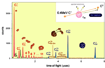
During collisions with fast heavy ions, C60 is highly excited and eventually decayed emitting many electrons and small fragment ions. By measuring these fragment ions with a time-of-flight coincidence method, quantitative understanding is achieved about anti-radiation stability and mechanisms leading to electronic excitations and how these excitations evolve with emission of secondary particles.
Ion-irradiation-induced polymerization of C60 thin film, energy and angular distribution of secondary fullerene ions
C60 thin film formed on a Si(111) substrate
is known to have a fcc crystal structure via weak van der Waals force.
The bond-strength of a film is in practice too weak to be used as a new
materials.
As a novel technique to overcome this problem, we employ the ion-irradiation
method using various slow-to-fast ions.
Also, we study a sputtering process resulting in the formation of large fullerenes such as C64 and (C60) 2. to understand the mechanism in detail, we investigate energy and angular distribution of these fullereren ions.
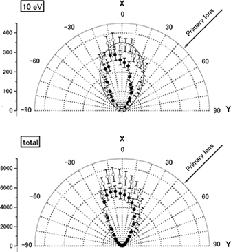
Angular and energy distribution of secondary ions produced from a C60 thin film bombarded by 4MeV Si ions.
Semiconductor Radiation Detectors
| 1) | Charge releasing radiation detector When boron-doped Si is cooled down below liquid He temperature, the borons can capture holes. The energy required to release a hole from boron is nearly 2meV, which is the same energy to break a cooper pair to make two quasi-particles in superconducting radiation detectors. This means that the cryogenic Si detector has the possibility of having the same energy resolution with superconducting detectors. To develop this type of radiation detector, we are fabricating metal-insulator-semiconductor device. |
| 2) | InSb compound semiconductor radiation detector To achieve high energy resolution in radiation detector, the energy to create a charge must be small. In Si, 3.6eV is necessary to produce a pair of electron-hole. However, in InSb, only 0.5eV is enough to have a pair of electron-hole. This implies that InSb will have 7 times as many as electron-hole pairs with Si. In other words, the energy resolution of InSb detector can be 2-3 times better than that of Si detector. One of the shortcomings of InSb detector is thermal noise at room temperature. We have to cool InSb detector down to certain temperature. We fabricated InSb radiation detector for the first time in the world, and measured the energy spectrum of alpha particles. |
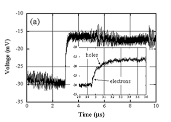
A pulse due to alpha particle measured by InSb radiation detector. Operating temperature is 0.5K.
Inset. Fast and slow rising parts are the contributions of electrons and holes, respectively.
X-ray transmission method with low dose exposure
Computed tomography (CT) is an useful tool to find tumors such as cancers. The dose exposure of CT is, however, 100-1000 times as much as that of breast radiograph.
Our purpose is to decrease the dose exposure of CT, and to employ CT as an annual health check. To meet this demand, we are developing high performance X-ray detector and high quality X-rays.
In case of detecting contrast media such as Iodine in a human body, we propose to utilize filtered X-rays and their energy information. With this system, the estimated dose exposure will be 40% of that of normal radiograph. We are also developing high efficiency, fast X-ray detector.
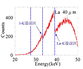
Energy spectrum of La filtered X-rays after passing 20cm of water layer with 100mm iodine contrast media.
We can observe the K-edges of La and I clearly.